

The human eye is an organ that reacts to light and has several purposes. As a sense organ, the mammalian eye allows vision. Rod and cone cells in the retina allow conscious light perception and vision including color differentiation and the perception of depth. The human eye can distinguish about 10 million colors.
Human eye Structure
Similar to the eyes of other mammals, the human eye’s non-image-forming photosensitive ganglion cells in the retina receive light signals which affect adjustment of the size of the pupil, regulation and suppression of the hormone melatonin and entrainment of the body clock.

HUMAN EYE
Size
The dimensions differ among adults by only one or two millimeters; it is remarkably consistent across different ethnicities. The vertical measure, generally less than the horizontal distance, is about 24 mm among adults, at birth about 16–17 millimeters (about 0.65 inch). The eyeball grows rapidly, increasing to 22.5–23 mm (approx. 0.89 in) by three years of age. By age 13, the eye attains its full size. The typical adult eye has an anterior to posterior diameter of 24 millimeters, a volume of six cubic centimeters (0.4 cu. in.), and a mass of 7.5 grams (weight of 0.25 oz.)
*Vision
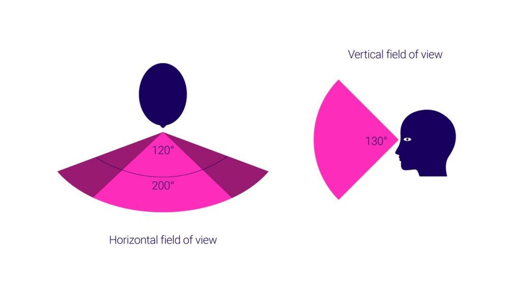
Field of view
The approximate field of view of an individual human eye is 95° away from the nose, 75° downward, 60° toward the nose, and 60° upward, allowing humans to have an almost 180-degree forward-facing horizontal field of view.
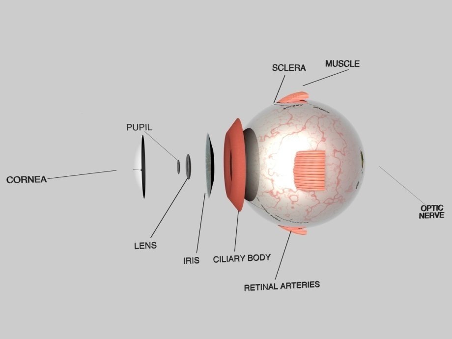
Parts of the eye.
Cornea
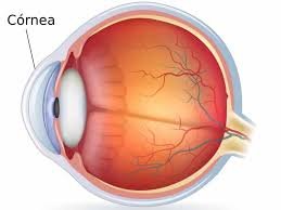
The cornea is the outer covering of the eye. This dome-shaped layer protects your eye from elements that could cause damage to the inner parts of the eye. There are several layers of the cornea, creating a tough layer that provides additional protection. These layers regenerate very quickly, helping the eye to eliminate damage more easily. The cornea also allows the eye to properly focus on light more effectively. Those who are having trouble focusing their eyes properly can have their corneas surgically reshaped to eliminate this problem.
Sclera
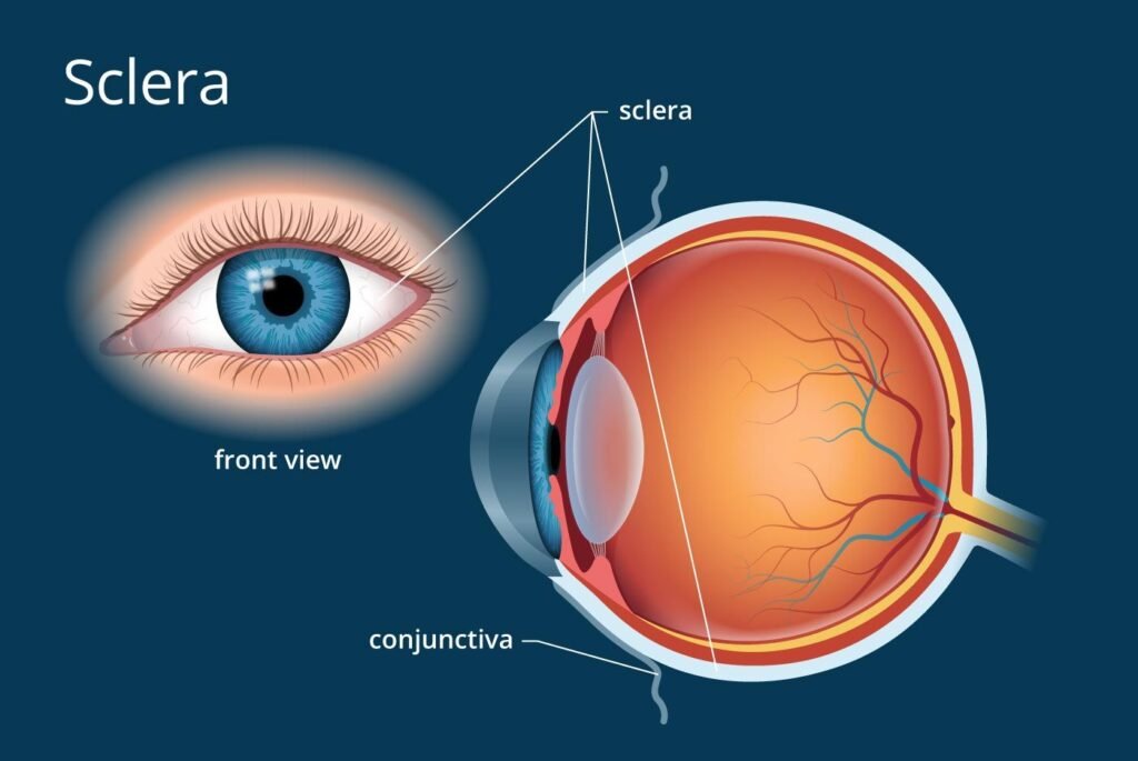
The sclera is commonly referred to as the “whites” of the eye. This is a smooth, white layer on the outside, but the inside is brown and contains grooves that help the tendons of the eye attach properly. The sclera provides structure and safety for the inner workings of the eye, but is also flexible so that the eye can move to seek out objects as necessary.
Pupil

The pupil appears as a black dot in the middle of the eye. This black area is actually a hole that takes in light so the eye can focus on the objects in front of it.
Iris
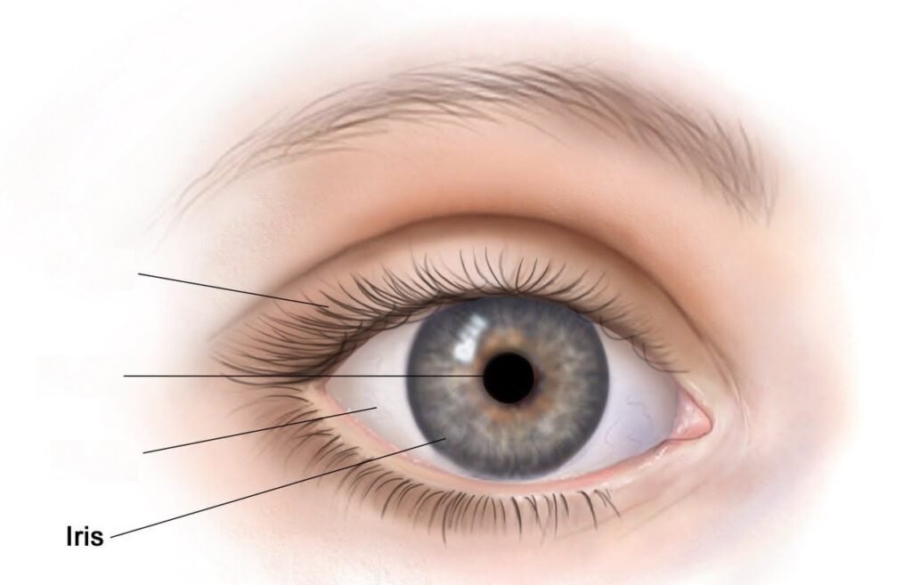
The iris is the area of the eye that contains the pigment which gives the eye its color. This area surrounds the pupil, and uses the dilator pupillae muscles to widen or close the pupil. This allows the eye to take in more or less light depending on how bright it is around you. If it is too bright, the iris will shrink the pupil so that they eye can focus more effectively.
Conjunctiva Glands
These are layers of mucus which help keep the outside of the eye moist. If the eye dries out it can become itchy and painful. It can also become more susceptible to damage or infection. If the conjunctiva glands become infected the patient will develop “pink eye.”
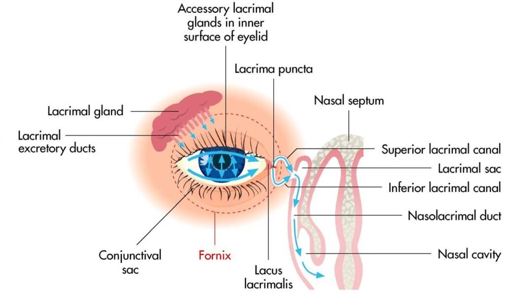
Lacrimal GlandsThese glands are located on the outer corner of each eye. They produce tears which help moisten the eye when it becomes dry, and flush out particles which irritate the eye. As tears flush out potentially dangerous irritants, it becomes easier to focus properly
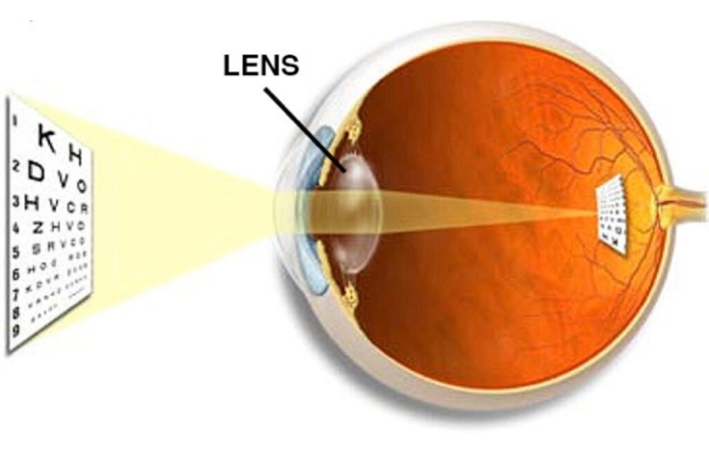
Lens
Retina
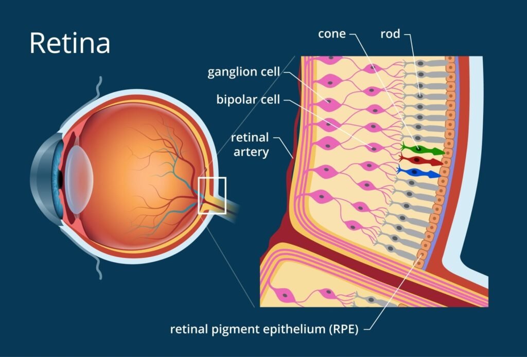
The light focuses by the lens will be transmitted onto the retina. This is made of rods and cones arranged in layers, which will transmit light into chemicals and electrical pulses. The retina is located in the back of the eye, and is connected to the optic nerves that will transmit the images the eye sees to the brain so they can be interpreted. The back of the retina, known as the macula, will help
Leave a Reply